
Figure 7.
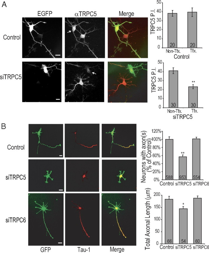
Regulation of axon formation by TRPC5 channels. A, E18 Hippocampal neurons were electroporated before plating with control siRNA or siTRPC5 plus soluble EGFP. Neurons were fixed at 48 h after electroporation and stained with anti-TRPC5 polyclonal antibody and imaged as described in Materials and Methods. Representative images from control siRNA (top) and siTRPC5 (bottom) are shown in left panel and the graphs summarizing quantification (y-axis shows TRPC5 P.I. in arbitrary units) are shown on right. B, E18 neurons were electroporated before plating with control siRNA, siTRPC5, or siTRPC6. Neurons were fixed at 48 h and evaluated for axon formation by staining with anti-Tau-1 antibody (see Materials and Methods). Representative images are shown in left panel(s) and quantification of percentage of polarized neurons (top graph) and length of (bottom graph) in the neurons that did polarize is shown on right. Scale bars: 20 μm (all panels).