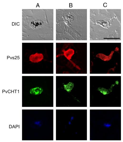
Fig. 2.
Immunofluorescence localization of P. vivax chitinase PvCHT1 in in vitro-developed mosquito midgut stage parasites. Column A (left), zygote; B, immature ookinete; C, mature ookinete. Each of 3 columns consists of 4 panels showing from top to bottom; differential interference contrast (DIC) image, staining with rabbit anti-Pvs25 antiserum (red), with mouse anti-PvCHT1 (green), and DAPI-stained nuclei (blue). Bar = 10 μm. See Section 2.6. (For interpretation of the references to color in this figure legend, the reader is referred to the web version of this article.)

