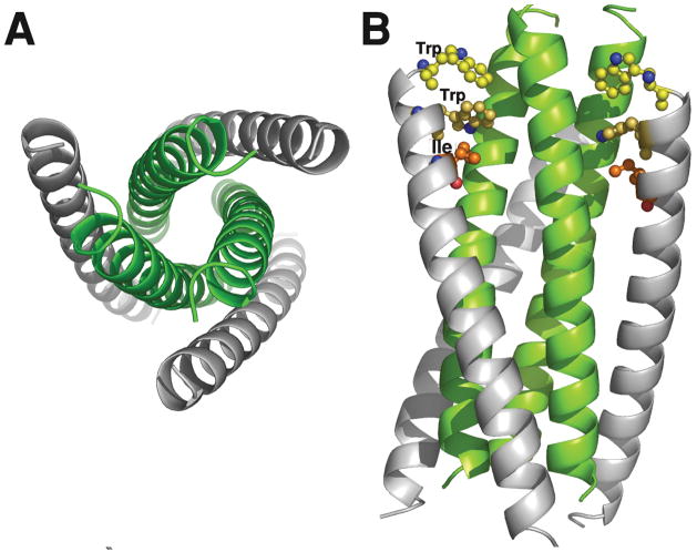
Figure 4.
X-ray structure of the gp41 6-helix bundle. The bundle was formed from synthetic N36 and C34 peptides (see Table 1). Panel A shows an end view of the 6-HB with the C-peptides in grey and the trimeric core in green. Panel B is a side view illustrating the trimeric N-core interacting with the C-peptides. The pocket binding domain (PDB; WMEWDREI) residues Trp, Trp and Ile of C34 are shown explicitly. These interact with the hydrophobic pocket of the N-trimeric core. The model was built using coordinates from the PDB 1AIK.

