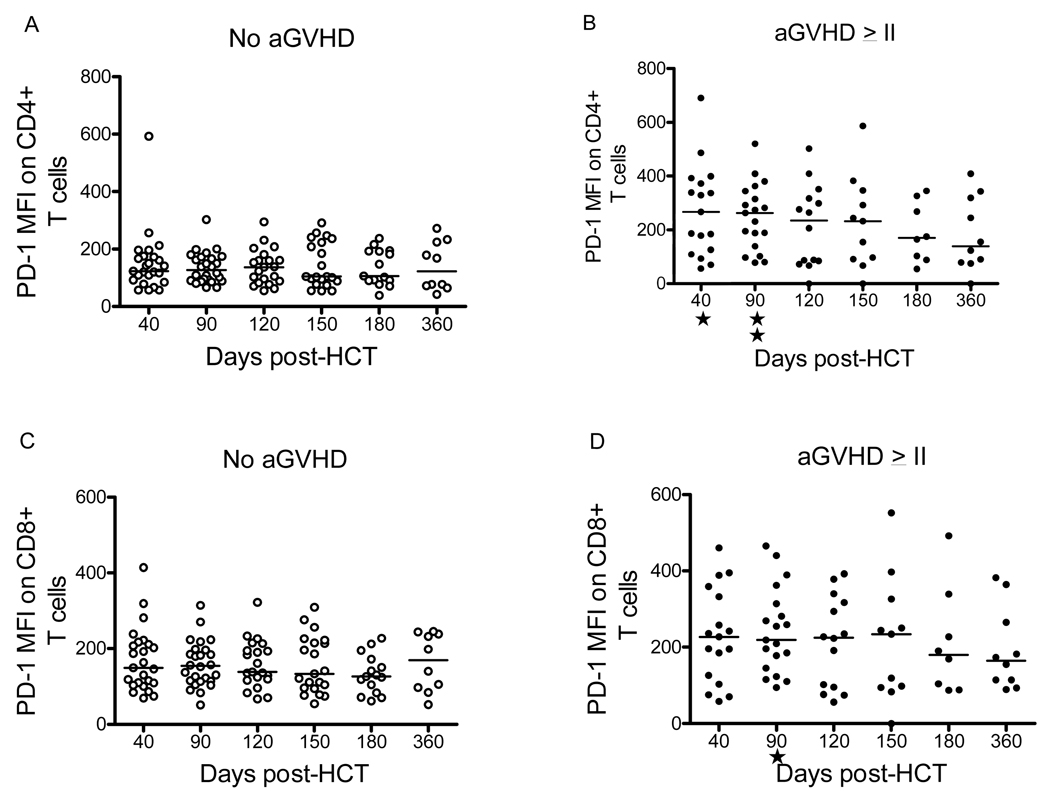
Figure 6. PD-1 Mean Fluorescence Intensive in CD4+ and CD8+ T cells of Non-viremic patients with or without GVHD.
Mean fluorescent intensity (MFI) is shown for patients with GVHD grade 0-I (panels A and C, labeled “No aGVHD”) or with GVHD >grade II (panels B and D, labeled “aGVHD >II”). Mann-whitney test was used to establish the association with MFI in CD4+/PD-1+ and h aGVHD >II (p= 0.01 at day 40 and p=0.001 at day 90) and between MFI of CD8+/PD-1+ cells and aGVHD >II on day 90 post-HCT (p= 0.01).

