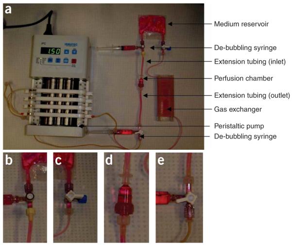
Figure 4.

A perfusion loop for cultivation of cardiac tissue constructs. (a) Assembled perfusion loop. Luer connectors are inserted into the tubing before the assembly. (b) Connection between the medium reservoir and the inlet extension tubing of the perfusion chamber. (c) Connection between the medium reservoir and the gas exchanger. The three-way stopcock is capped with a male luer cap. (d) Connection between the perfusion chamber and the inlet/outlet extension tubing. (e) Connection between the outlet extension tubing of the perfusion chamber and the pump tubing.
