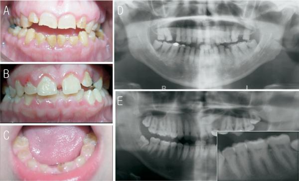
Figure 2.
Clinical photographs and radiographs. (A) Frontal view of affected individual II-2 at age 39. Note mild discoloration with minor attritions. (B) Frontal view of affected individual III-1 at age 15. Clinically permanent teeth look normal. (C) Frontal view of affected individual III-4 at age 4. Note brown discoloration and attrition. (D) Panoramic radiograph of affected individual II-2 at age 39. Note almost complete pulpal obliterations. (E) Panoramic radiograph of affected individual III-1 at age 15. The premolar and canine teeth have thistle-shaped pulp chambers with pulp stones. The inset is a magnified view of left mandibular premolars and molars. Note thistle-shaped pulp chambers and pulp stones.

