
Figure 2.
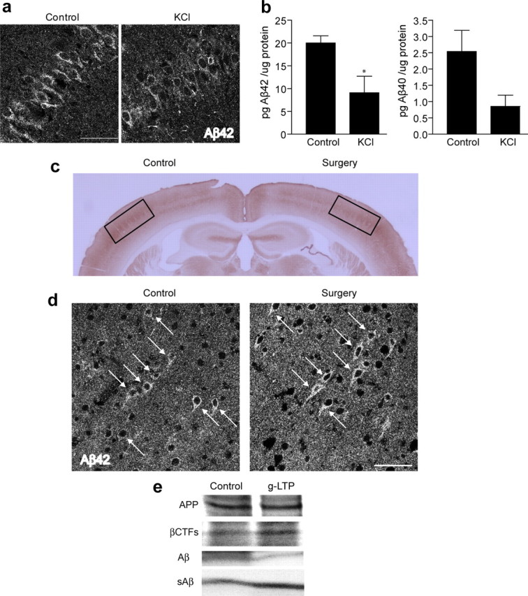
Synaptic activation reduces intraneuronal Aβ in hippocampal slices, whereas chronic inhibition of synaptic activity induces intraneuronal Aβ accumulation in vivo. a, Hippocampal slices prepared from Tg19959 mice and incubated for 8 h with KCl. Confocal microscopy of CA1 neurons demonstrates 13 ± 2% reduction of intraneuronal Aβ42 immunofluorescence in KCl-treated compared with control slices (n = 3; p < 0.01). b, Aβ1-42 ELISA of lysates from hippocampal slices treated with KCl versus control (n = 3; p < 0.05); Aβ1-40 ELISA of lysates from slices treated with KCl versus control (n = 3; p = 0.09). c, Representative section of Tg19959 brain stained for COX. The side corresponding to whisker removal (Surgery) shows a dramatic decrease of COX compared with the control side (Control). d, Confocal microscopy showing intraneuronal Aβ42 immunofluorescence of Tg19959 barrel cortex. The neuron cell bodies corresponding to the side with chronic synaptic inhibition (Surgery) show 10 ± 1% higher levels of Aβ42 compared with neurons on the control side (n = 4; p < 0.05). e, Metabolic labeling revealed decreased levels of newly generated Aβ (43 ± 11% decrease; n = 4; p < 0.01) and increased levels of newly generated βCTFs (22 ± 8% increase; n = 4; p < 0.05), in the presence compared with the absence of g-LTP. Aβ and βCTF values were normalized to newly generated APP. Levels of newly generated secreted Aβ (sAβ) (normalized to newly generated APP) were increased by 44 ± 15% in media of g-LTP activated compared with control Tg2576 neurons (n = 4; p < 0.05). Scale bars, 50 μm.