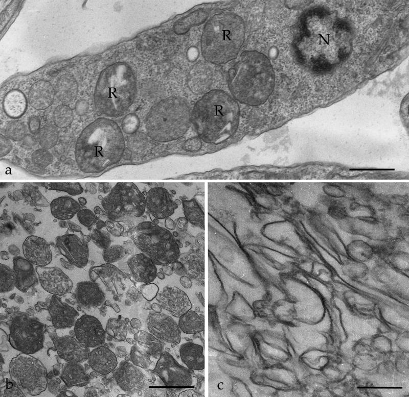
Figure 1.
Transmission electron microscopy of T. cruzi reservosomes. (a) Ultrathin section of an epimastigote showing reservosomes (R) in situ, with their typical morphology and position, between nucleus (N) and posterior end of the cell. (b) Purified reservosome fraction (B1). (c) subcellular fraction containing reservosome membranes (B1M). Bars represent 0.3 μm (a) and 1 μm (b,c).

