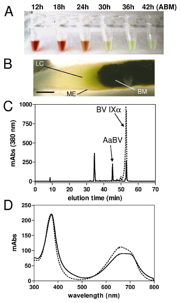
Figure 1.

Accumulation of a bilin pigment in the midgut of A. aegypti after a blood meal. (A) Midgut dissected at different times after a blood meal (ABM) were homogenized in PBS and centrifuged. Tubes containing the supernatants are shown. (B) A. aegypti midgut 38 h ABM. Midgut epithelium (ME); lumenal content (LC); blood meal (BM); scale bar = 0.2 mm. (C) A. aegypti pigment was purified by reverse phase HPLC using a C18 column. Chromatograms of A. aegypti midgut PBS extract (solid lines) and standard biliverdin IXα (dashed lines) are shown. (D) Light-absorption spectra of peaks containing AaBV (solid line) and BV IXα (dashed lines).
