
Figure 3. Comparison of the properties of redox modulation, permeability for Ca2+ versus Ba2+, and Ni2+ inhibition of T-type Ca2+ currents among three subgroups of T-rich cone bipolar cells.
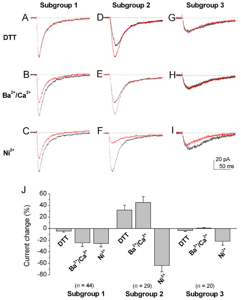
Currents were evoked by a voltage step pulse to -40 mV from a holding potential of -80 mV. Three subgroups of cone bipolar cells were observed based on the extent to which their T-type Ca2+ currents were sensitive to modulation by 2 mM DTT, were permeable to Ca2+ versus Ba2+, and were sensitive to 20 μM Ni2+. (A-C) The T-type Ca2+ currents in the first subgroup did not show significant DTT modulation (A), they were less permeable to Ba2+ than to Ca2+ (B) and they showed low sensitivity to Ni2+ inhibition (C). (D-F) The T-type Ca2+ currents in the second subgroup were potentiated by DTT (D). In addition, they were more permeable to Ba2+ than to Ca2+ (E), and they were more sensitive to Ni2+ inhibition (F). (G-I) The T-type currents in the third subgroup did not show significant modulation by DTT, they were equally permeable to Ca2+ and Ba2+, and they had low sensitivity to Ni2+ inhibition. (J) The bar graphs are the sums of the percentage changes to control of T-type currents in DTT modulation, permeability to Ca2+ versus Ba2+, and Ni2+ inhibition in three subgroups of cone bipolar cells. The T-type currents in the second subgroup of cone bipolar cells were significantly more sensitive to Ni2+ inhibition than those of the other two subgroups (p < 0.05). Error bars indicate the SD.