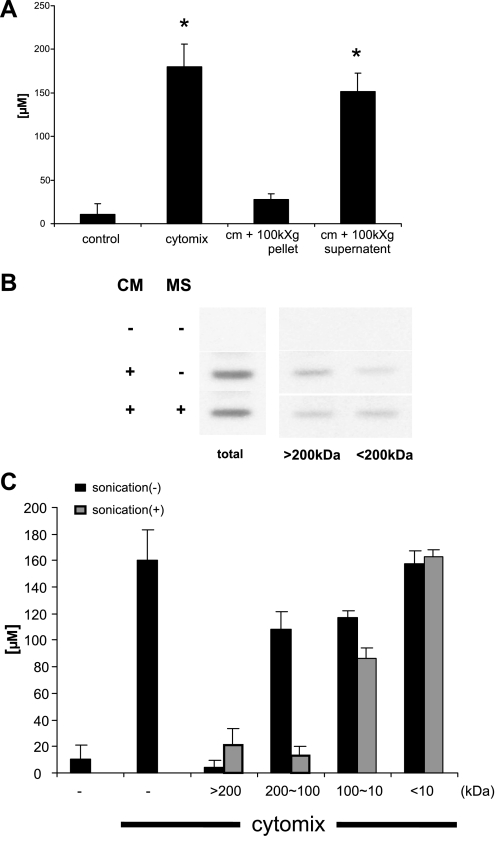
Fig. 4.
The iNOS-suppressing factor has an apparent molecular mass between 100 and 200 kDa and is present in the microsomal compartment of LC. The microsomal compartment was isolated from LC as described in materials and methods. Immunostimulated Caco-2 cells were exposed to either resuspended microsomes or the postmicrosomal supernatant for 24 h. A: NO2−/NO3− concentration was measured by Griess assay. B: inhibition of iNOS dimerization by the resuspended microsomes (MS) was performed as described in Fig. 3. C: 200 μl of LC was diluted to 1 ml with isotonic Tris buffer (25 mM Tris, pH 7.4, 130 mM NaCl) and was or was not sonicated for 30 s on ice and then spun at 15,000 g for 10 min. The supernatant was filtered sequentially through MWCO of 200-, 100-, and 10-kDa membranes. Each retentate was reconstituted to the original volume with Tris buffer and then added to immunostimulated Caco-2 cells, and NO2−/NO3− concentration was measured by Griess assay. *P < 0.05 vs. control.

