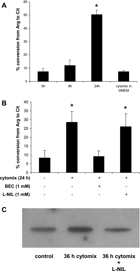
Fig. 6.
Arginase in liver cytosol is activated by supernatant of immunostimulated Caco-2 cells. A: Caco-2 cells were treated with vehicle alone or cytomix for 18 h, 500 μl of each supernatant was harvested and centrifuged at 1,000 g for 10 min to remove cell debris; 2 μl of LC (20 mg/ml) was added to 25 μl of each supernatant. After incubation for the times indicated in the figure the mixture was subjected to the arginase activity assay as described in materials and methods. As a negative control cytomix was added to 2 μl of LC in 25 μl of DMEM and subjected to the arginase activity assay. B: in the same system, BEC (1 mM) and l-N6-(1-iminoethyl)-lysine·2HCl (l-NIL; 700 μM) were added, respectively, to be subjected to the urea assay as described in the same way as in Fig. 5D. C: to investigate S-nitrosylation of the Arg-1 in LC, LC was subjected to biotin switch assay as described in materials and methods. *P < 0.05 vs. control.

