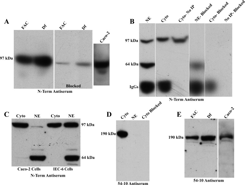
Fig. 10.
Western blot analysis and immunoprecipitation (IP) of Atp7a protein in IEC-6 and Caco-2 Cells. Western blot analysis of membrane proteins is shown (N-term; A). IP analysis of IEC-6 cell proteins from cytosolic (Cyto) and nuclear extracts (NE) was performed (B). Western blot of cytosolic and nuclear fractions from Caco-2 and IEC-6 cells is shown in C (N-term). Western blotting studies with the 54–10 antiserum are depicted in D and E. Representative experiments are shown.

