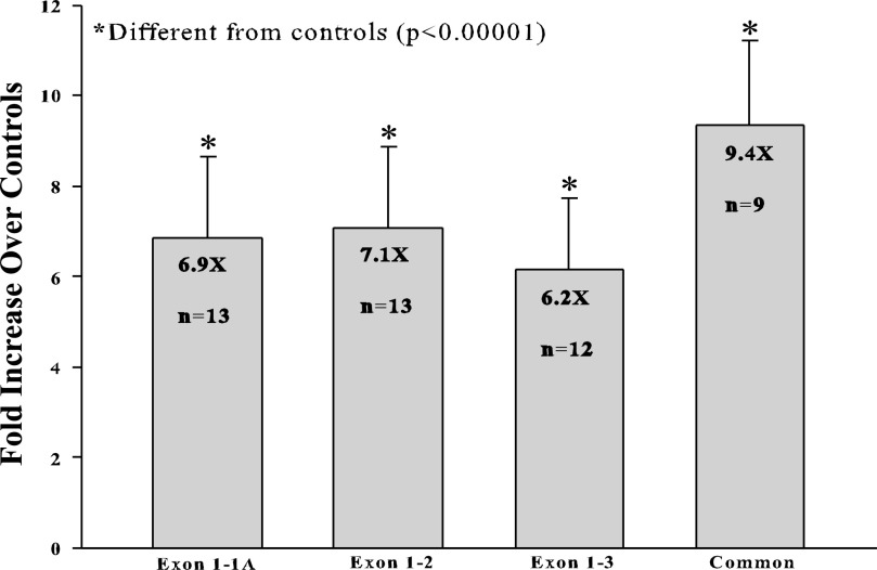
Fig. 5.
qRT-PCR analysis of Atp7a splice variants in rat intestine. Several different primer sets designed to detect the different splice variants were used to quantify Atp7a mRNA expression in the proximal intestines of control and iron-deficient rats. Exon 1–1A, forward primer in exon 1 and reverse primer in exon 1A; Exon 1–2, forward primer in exon 1 and reverse primer that spans the junction between exons 1 and 2; Exon 1–3, forward primer in exon 1 and reverse primer that spans the junction between exons 1 and 3; Common, primers that bind to exons 6–7 (downstream of the region where alternative splicing occurs) and presumably amplify all Atp7a transcript variants. Fold inductions and n values are shown in each bar.

