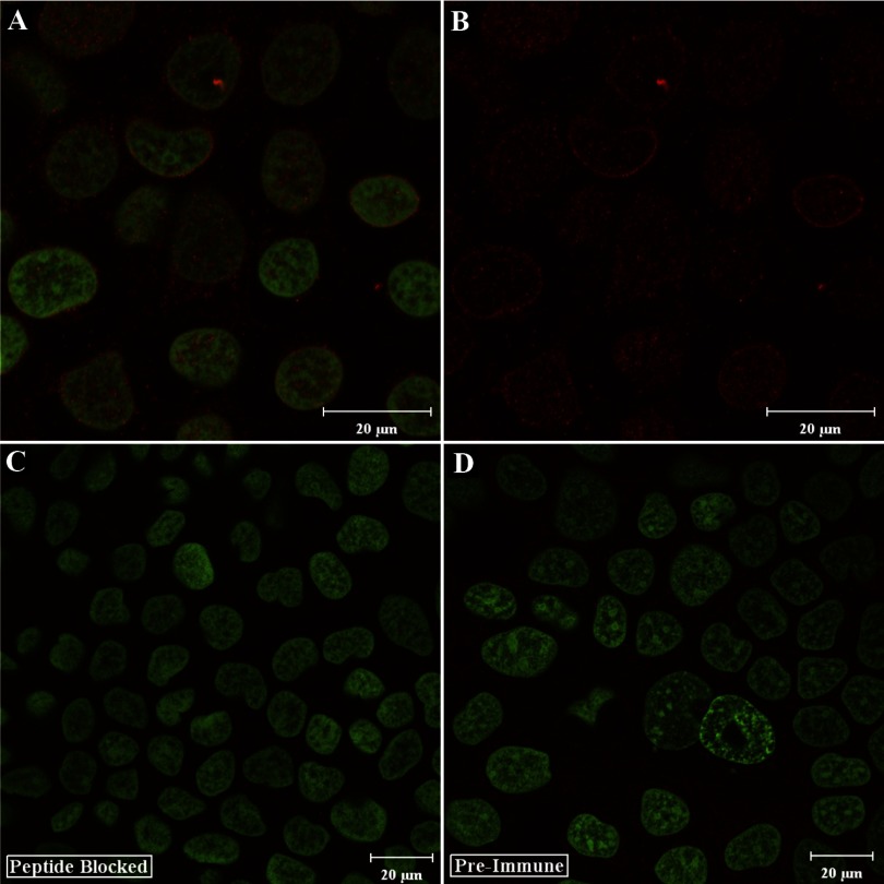
Fig. 8.
Immunocytochemical analysis of Atp7a protein expression in differentiated Caco-2 cells using the N-term antibodies. Caco-2 cells were grown to 20 days postconfluence and then reacted with the N-term antibodies followed by staining with Sytox Green. A: overlay of the 647 channel (Atp7a; red color) and the 488 channel (Sytox Green). B: only the Atp7a staining. Peptide-blocked antibodies or preimmune serum were used in some experiments, as seen in C and D (which show the overlay of the 647 and 488 channels). Typical images are shown from one of several experiments.

