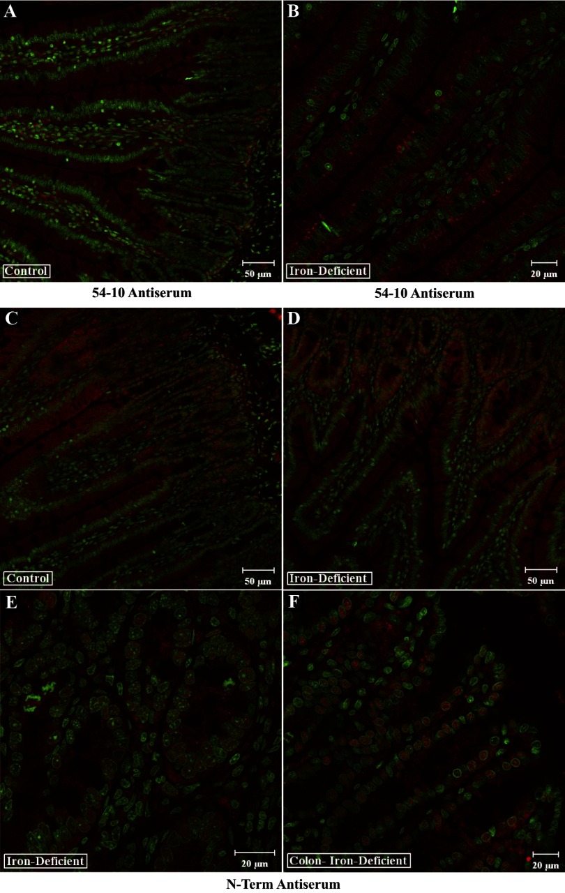
Fig. 9.
Immunohistochemical analysis of rat intestine with Atp7a antibodies. Transverse sections of intestinal tissue were reacted with Atp7a antibodies followed by staining with Sytox Green. Shown in all panels are overlays of the 647-nm laser line (detecting Atp7a; red color) and the 488-nm laser line (detecting Sytox Green). The 54–10 antiserum was used with sections from control and iron-deficient rats (A and B). The N-term antibodies were also utilized in parallel experiments with samples derived from small (C and D) and large (E and F) intestine. Typical images are shown that are representative of several experiments that were performed.

