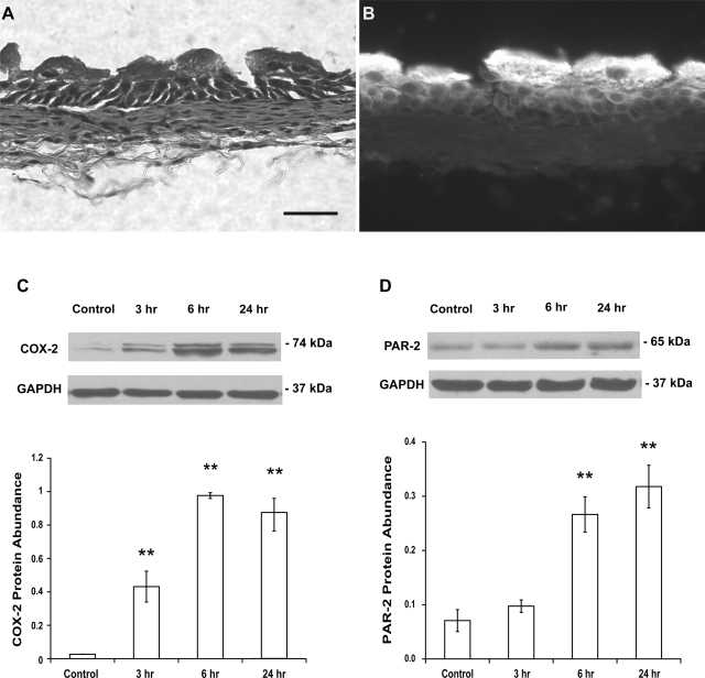
Fig. 1.
A: histological appearance (hematoxylin and eosin staining) of urothelium/suburothelium reveals that the contamination of detrusor was very limited. Scale bar, 50 μm. B: strong staining with a specific uroplakin antibody is shown in superficial urothelial cells. C: treatment of mice with cyclophosphamide (CYP; 150 mg/kg ip) for 3, 6, and 24 h induced an increase in cyclooxygenase-2 (COX-2) protein abundance in the bladder urothelium/suburothelium, respectively. D: similarly, treatment of mice with CYP induced an increase (6 and 24 h) in protease-activated receptor-2 (PAR-2) protein abundance in the bladder urothelium/suburothelium. Control mice received saline (ip). Relative COX-2 or PAR-2 protein abundance was analyzed by immunoblotting. Values determined by densitometry were normalized to those of GAPDH (loading control) in each sample. Data are means ± SE; n = 4–5. **P < 0.01 vs. control.

