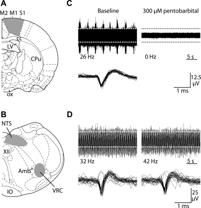
Fig. 1.
Multichannel recordings of spontaneous neuronal activity in cortical and medullary slices from interbout aroused (IBA) ground squirrels. A: cortical activity was recorded from neurons in the primary motor (M1), supplementary motor (M2), and primary somatosensory (S1) areas. Slices were cut approximately −1.30 mm caudal to bregma [anatomical images adapted from Paxinos and Watson (50)]. Shaded areas indicate electrode placement. B: medullary activity was recorded from the nucleus tractus solitarius (NTS) and ventral respiratory column (VRC). Slices were cut approximately −13.68 mm caudal to bregma. Shaded areas indicate electrode placement. C: representative cortical recordings are shown during baseline (top left) and after 60 min of pentobarbital sodium (300 μM) treatment (top right). Traces that cross the detection threshold (dashed white lines) are overlaid (bottom right). D: representative recordings from VRC neurons are shown during baseline (top left) and after 60 min of sodium pentobarbital (300 μM, top right). Traces that cross the detection threshold (dashed gray lines) are overlaid (bottom right).

