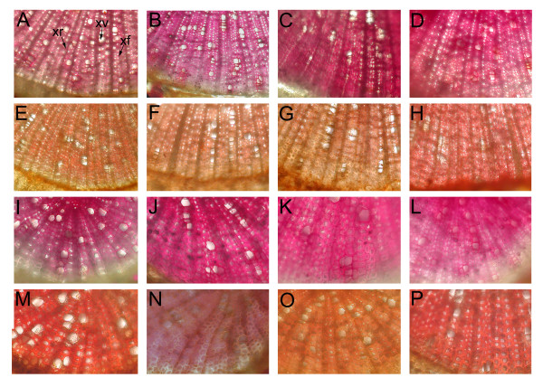
Figure 3.
Histochemical localization of lignin in non-inoculated silver birches. Cross-sections of stem and root of non-inoculated clone A (A, E, I, M) and PtCOMT-modified lines 23 (B, F, J, N), 44 (C, G, K, O) and 65 (D, H, L, P). Stems (A-H) and roots (I-P). Lignin stained pink-red in the phloroglucinol-HCL stained sections (A-D, I-L). In the Mäule stainings (E-H, M-P), syringyl lignin pink-red and guaiacyl lignin light brown to dark brown. xf, xylem fibre; xr, xylem ray; xv, xylem vessel.

