Figure 3.
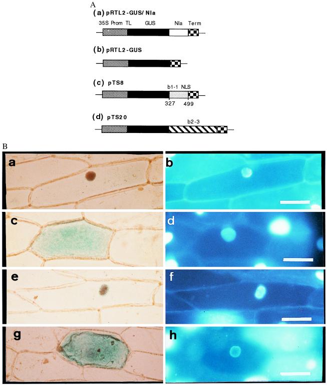
Subcellular localization of GUS activity directed by sequences from the A mating type proteins of C. cinereus. (A) Constructs introduced into onion cells. Coding regions were expressed under the control of the cauliflower mosaic 35S promoter and nopaline synthase terminator. (a) Control in which GUS was fused to the plant potyviral Nla protein that contains a bipartite NLS. (b) Control with GUS sequence alone. (c) GUS fused to amino acids 327–499 from b1–1. (d) GUS fused to b2–3. (B) GUS activity (a, c, e, and g) and 4′,6-diamidino-2-phenylindole-stained nuclei (b, d, f, and h) in transiently transformed onion epidermal cells. GUS activity localized to the nucleus by the Nla protein (a and b), GUS alone localized to the cytoplasm, (c and d) GUS activity localized to the nucleus by the predicted b1–1 NLSs (e and f), and GUS activity localized to the cytoplasm when fused to b2–3 (g and h). (Bar = 100 μm.)

