Fig. 3.
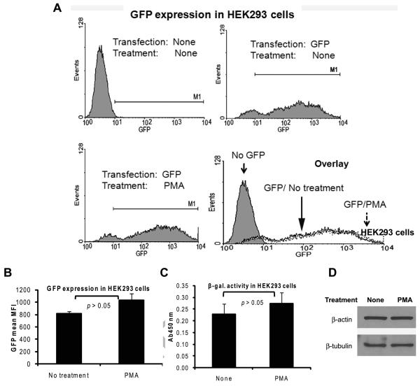
The induction of Renilla luciferase expression by PMA is not due to a global increase in gene expression in HEK293 cells. A and B: HEK293 cells transfected with 200 ng of a plasmid expressing GFP under the CMV promoter were left untreated or treated with PMA for 8 hours beginning at 24 hours post-transfection. The cells were then harvested and flow cytometry was carried out with excitation and emission at 488 nm and 530 nm, respectively. Representative histograms of live cells gated based on light scatter profiles are shown in panel A. The upper left histogram is from cells transfected with an empty vector plasmid. The mean and SD of the MFI of the GFP signal in the GFP-positive cells from three independent experiments are shown in panel B. C: HEK293 cells transfected with 100 ng of a plasmid expressing β-galactosidase under the SV40 promoter were left non-treated or were treated with PMA for 8 hours. The cells were then harvested β-galactosidase assay was performed. The results show the mean and SD of three independent experiments. D: Overnight cultures of HEK293 cells were left non-treated or were treated with PMA for 8 hours. At the end of the treatment, the cells were harvested and immunoblotting was performed for β-actin and β-tubulin.

