Fig. 4.
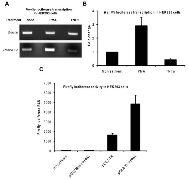
PMA induces Renilla luciferase expression by stimulating the HSV-1 TK promoter. A and B: HEK293 cells grown on 100-mm plates were transfected with 120 ng of pRL-TK plasmid and they were then left non-treated or treated with PMA or TNFα for 5 hours beginning at 24 hours after transfection. Total RNA was isolated and duplex RT-PCR was carried out on 100 ng of total RNA using primers specific for Renilla luciferase and β-actin. The PCR products were resolved by agarose gel electrophoresis. A representative image of the bands is shown in panel A. The mean and SD of the band intensities of Renilla luciferase bands normalized to those of the corresponding β-actin bands from three independent experiments are shown in panel B; fold change was calculated by setting the value for the non-treated samples as 1. C: HEK293 cells transfected with 100 ng of either the pGL2 Basic or the pGL2-TK plasmid were left non-treated or treated with PMA for 8 hours beginning at 24 hours post-transfection. The cells were then harvested and firefly luciferase assays were performed. The results show the mean and SD of the normalized RLU values from three independent experiments.

