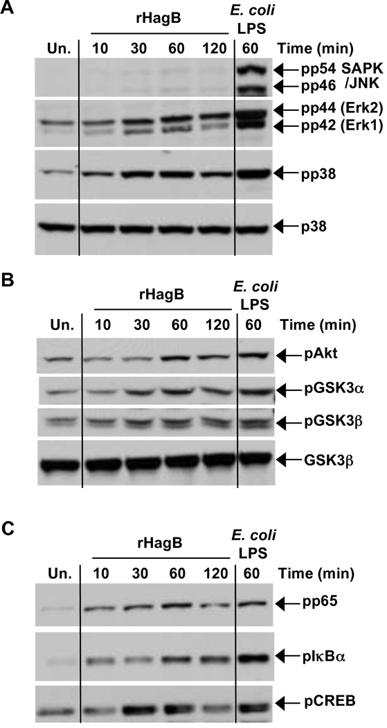
Fig. 2.
Signaling pathways activated following rHagB stimulation of DC. DC were stimulated with 40 µg/ml rHagB for 10, 30, 60 or 120 min. Following stimulation, cells were lysed and whole cell lysates were assessed for phosphorylation of (A) JNK, ERK1/2 and p38, (B) Akt and GSK3α/β and (C) NF-κBp65, IκBα and CREB by Western blot. Total p38 (A) and GSK3β (B) were used as loading controls. Unstimulated DC (far left lane) or DC stimulated with 100 ng/ml E. coli K12 LPS for 60 min (far right lane) were used as controls. Results represent one of three independent experiments.

