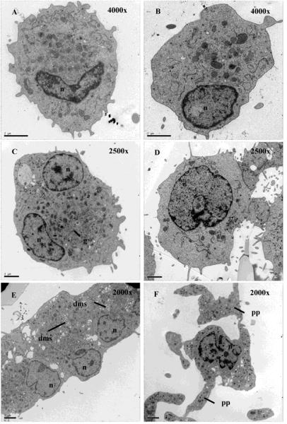
Figure 1.
Mks cultured with NIC exhibit normal ultrastructure. Transmission electron microscopy of Mks cultured with Tpo only (A-B) and Tpo + NIC (C-F) at day 8. The images in A-D were acquired such that the magnification gave one cell per field of view. Due to the larger cell size with Tpo + NIC, it was necessary to use a lower magnification (2500x) compared to that for Tpo only (4000x). (E) An image of 3 cells exhibiting a demarcation membrane system (2000x). (F) A proplatelet-bearing Mk (2000x). n = nucleus, g = granules, dms = demarcation membrane system, pp = proplatelet. Scale bar = 2 μm.

