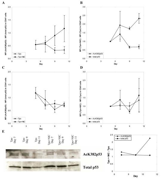
Figure 5.
NIC increases acetylation of p53. mPB CD34+ cells were cultured with 100 ng/mL Tpo. On day 5 cultures were supplemented with 6.25 mM NIC. (A-D) Intracellular flow cytometry was used to detect expression of AcK382p53 and total p53 in (A-B) CD41+ and (C-D) CD41- cells. Mean fluorescence intensity (MFI) was calculated by subtracting the MFI of an unstained sample from the MFI of a stained sample to correct for background fluorescence. Data shown represent the mean ± SEM of two experiments. (A,C) The MFI of cells stained with an antibody against AcK382p53 was divided by the MFI of cells stained with an antibody against total p53. This ratio is shown for cells maintained in Tpo with or without NIC. (B,D) The MFI of cells grown with NIC was divided by the MFI of cells grown with Tpo only. This ratio was calculated for cells stained either with an antibody against AcK382p53 or against total p53. Based on a paired t-test, values of p < 0.1 (*) are indicated for the various time points. (E) Nuclear lysates of unselected cells were prepared at all time points shown and loaded onto SDS-PAGE gels. After electrophoresis, the proteins were transferred to PVDF membranes for Western blots. After probing for AcK382p53 residues, the blots were stripped and probed for total p53. Corresponding densitometry analysis is given. The blot shown is representative of two biological experiments.

