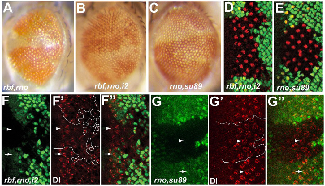
Figure 6.
Removal of the transcription activation domain of dE2F1 suppressed differentiation defects in rbf,rno mutant clones. (A–C) Adult eye images of rbf,rno clones in WT (A), rbf,rno clones in de2f1i2/rm729 (B), and rno clones de2f1su89 (C) background. (D–E) Anti Sens staining (shown in red) revealed low incidence of multiple-R8 phenotypes in rbf,rno clones in the de2f1i2/rm729 background (D) and in rno clones in the de2f1su89 background (E). (F–G”) Anti Dl antibody staining of Dl protein (red) in rbf,rno clones in the de2f1i2/rm729 (F–F”) and rno clones in de2f1su89 (G–G”) backgrounds are shown. Mutant clones were marked by absence of GFP and are outlined in F’ and G’. White arrows and arrowheads point to Dl protein expression in WT and mutant clones, respectively.

