
Figure 2.
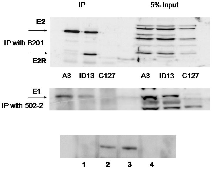
A3 cells, ID13 cells and the parental cell line C127, were lysed and immunoprecipitated with monoclonal antibody to BPV-1 E2 (B201). The proteins were immunoblotted using the rabbit antibody II-1 and visualized with the Thermo-scientific dura ECL detection kit (top panel). A3, ID13 and C127 cells were blocked overnight in 5 mM thymidine and BPV-1 E1 was immunoprecipitated using the 502-2 rabbit antibody. E1 was then detected with 502-2 (center panel). A3 cells were blocked in thymidine (lane 2), uncycled (lane 3) or blocked overnight with nocodazole (lane 4) and immunoprecipitated and blotted with the 502-2 rabbit antibody. Lane 1 shows thymidine treated C127 cells (bottom panel).