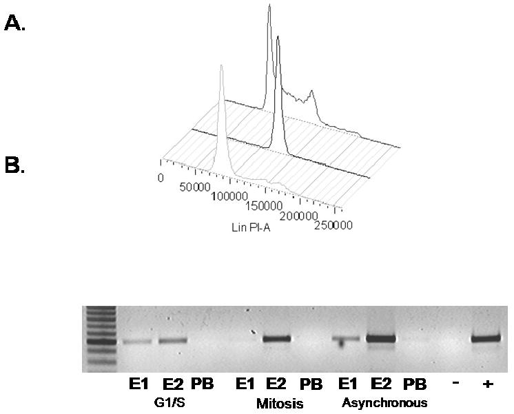
Figure 4.

A3 cells were synchronized using either 5mM thymidine to block in G1/S (forefront), 100 ng/ml nocodazole to block in mitosis (center plot), or were left untreated (rear plot), for 12 to 16 hours then harvested and stained with propidium iodide for FACS (A). Cycled cells were harvested and ChIP assays performed using E1 antibody 502-2, the E2 antibody II-1 or rabbit pre-immune serum (B).
