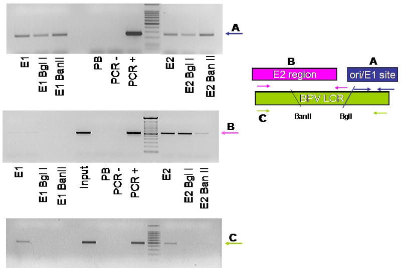
Figure 5.

A3 cells were synchronized and harvested for ChIP assay. Lysates were digested with either Bgl I or Ban II then immunoprecipitated with 502-2, II-1 or pre-immune sera. (A) PCR reactions were performed for the BPV origin of replication; (B) the region of the LCR immediately upstream of the origin, or (C) spanning the origin and upstream regions. The figure on the right is a schematic representation of primer usage with A, B and C representing primer pairs and amplified regions as described in Materials and methods.
