
Figure 7.
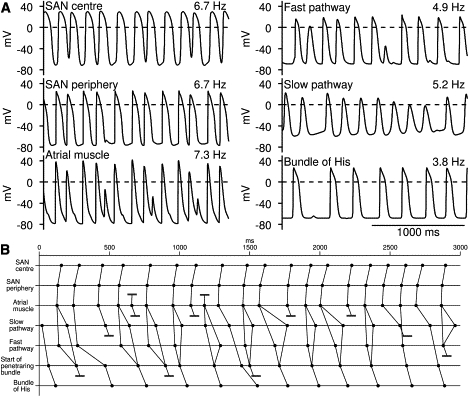
Protecting ventricles during atrial fibrillation. In this simulation, 1D multicellular model shown in Fig. 3A was used. To simulate AF, middle of string of atrial cells was stimulated and stimulus interval was changed randomly from 75 to 150 ms. (A) Selected action potentials. (B) Activation times at center and periphery of SAN, middle of atrial muscle, middle of fast and slow pathways, start of penetrating bundle, and bundle of His (middle of penetrating bundle). Conduction block is indicated by pair of parallel lines.