
Figure 9.
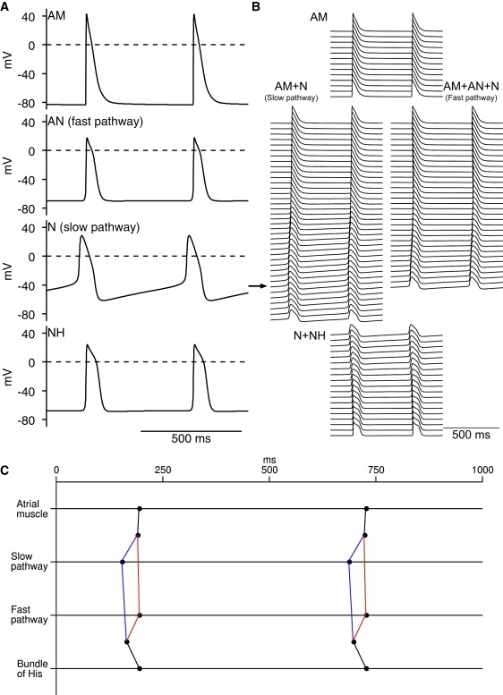
Pacemaker activity in 1D multicellular model of AVN. Multicellular model shown in Fig. 5A was used; it did not include SAN. (A) Selected action potentials. (B) Action potentials from many cells along length of multicellular model. (C) Activation times at middle of atrial muscle, middle of fast and slow pathways and bundle of His (middle of penetrating bundle). In absence of stimulation, AVN acted as pacemaker. Horizontal arrow in B indicates leading pacemaker site in slow pathway. Action potential propagated from leading pacemaker site in slow pathway to atrial muscle and NH cells; in addition, action potential propagated to center of fast pathway from both ends of fast pathway. AM, atrial muscle.