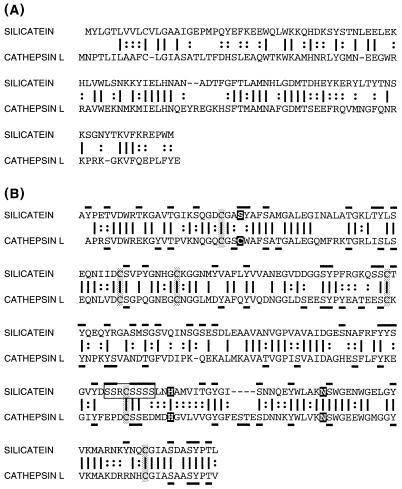
Figure 5.
Alignment and comparison of silicatein α with human cathepsin L. (A) Propeptide regions. (B) Mature proteins. Identical amino acids are indicated by vertical bars; similar residues indicated by colons. Cysteine residues involved in disulfide bonds in cathepsin L are shaded. Catalytic triad amino acids of the active site of cathepsin L and corresponding amino acids in silicatein α are highlighted. Hydroxy amino acid residues in silicatein α and cathepsin L are overlined and underlined, respectively. Silicatein α-specific hydroxy amino acid cluster is boxed.

