
Fig. 5.
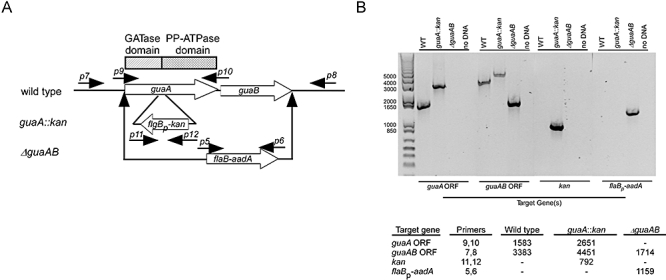
Characterization of guaA::kan and ΔguaAB mutant strains. A. Diagram of gene organization for wild-type guaAB, as well as the insertion points of antibiotic-resistance cassettes for the two mutant strain constructs (guaA::kan and ΔguaAB). The positions of the primers used for PCR are identified by number and correspond to those listed in Table 3. The location of GATase and PP-ATPase catalytic domains in guaA are indicated by rectangular boxes. B. Agarose gel containing PCR products using genomic DNA from the specified B. burgdorferi strains and primer sets corresponding to the target gene of interest. Standards in base pairs are shown on the left. Predicted product size (in base pairs) for each primer pair is indicated in the inset table.