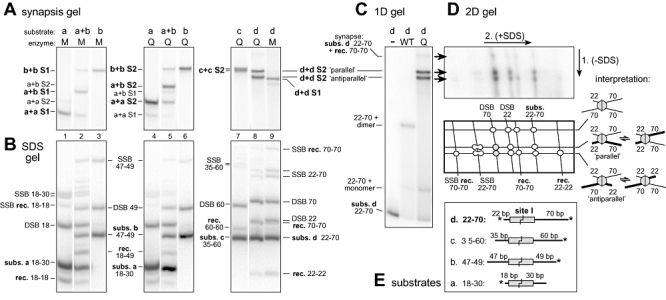
Fig. 6.

Stabilization of catalytically active site I synaptic tetramers. A. Separation of site I synaptic tetramers formed in vitro by the activated mutants Q115R and T77I/I100T/Q115R (Q and M respectively), by non-denaturing PAGE. The site I substrates a–d have different arm lengths, as depicted in (E). The synapse species designated S1 and S2 are named according to their component DNA substrates (e.g. b+b S2 contains two equivalents of substrate b). Reactions (24°C, 60 min) contained ∼12.5 nM site I with 125 nM Sin subunits. Only the part of the gel containing the synapses is shown (see C for a complete gel) and the lanes have been rearranged. Note that recombination intermediates present in the reaction mixtures (with mutant M in particular) may not all have been trapped as stable synapse intermediates visible on this gel (data not shown). B. Denaturing (+ SDS) gel analysis of samples from the same reactions as (A). Recombinant product and non-recombinant substrate DNA fragments (rec. and subs.) are indicated. The single-strand break (SSB) and double-strand break (DSB) products all have one Sin subunit covalently attached to the DNA, and are identified according to the attached DNA fragment. Note that when the DNA arms flanking site I are of sufficiently different lengths, the top and bottom strand SSB products migrate as a doublet of bands of equal intensity. C. Non-denaturing ‘synapsis’ gel. The Q115R (Q) reaction is similar to that in lane 8 of the gel in (A), but with ∼80 nM substrate d and 500 nM Q115R Sin. Marker reactions used ∼12 nM substrate with enzyme dilution buffer (−) or 250 nM WT Sin. Note that the synapse labelled ‘subs. d 22-70 + rec. 70-70’ is a very minor species and has the mobility expected for a synapse containing three 70 bp half-sites and one 22 bp half-site; its rarity suggests that very little dissociation/reassociation of synapses occurs during the reaction. D. Two-dimensional gel analysis of an aliquot of the sample run in (C). The first dimension is non-denaturing, and the second dimension is denaturing (+ SDS), as indicated. An interpretation of the spots, and cartoons of the presumptive synaptic complexes, are also shown. E. Structures of the site I substrates a–d. Substrates were 3′ end-labelled as shown (*); note that in the 22–70 substrate, the specific activities of the 70 and 22 ends are in the ratio ∼2:1.
