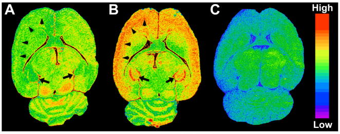
Figure 2.
Autoradiographic images of [125I]iodoDPA-713 [125I]1 to TSPO in the rat brain. Representative horizontal images of [125I]1 binding to TSPO in a normal rat brain (A) and in a neurotoxicant-injected rat brain (B). There is a significant increase in TSPO levels in the cerebral cortex (arrowheads) and hippocampus (arrows) in the neurotoxicant-injected rat brain (B) relative to the control brain (A). The image in (C) is representative of [125I]1 non-specific binding using 10 μM R-PK11195 as the blocking agent in a neurotoxicant-treated rat.

