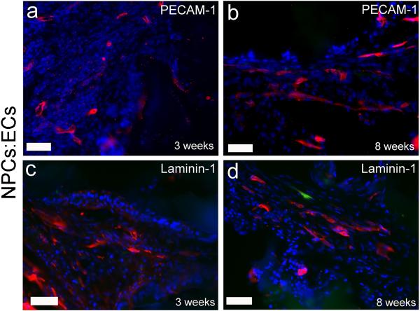
Figure 4. PECAM-1 and laminin-1 positive vessels at early and late time points.
Representative images of cross-sections from an implant plus NPCs:ECs group immunostained for PECAM-1 and laminin-1. Similar morphologies were seen for all groups. All images are from the lesioned side at the injury epicenter. (a) PECAM-1 (red) at three weeks and (b) eight weeks. Laminin-1 (red) staining at (c) three weeks and (d) eight weeks. The green cell in (d) is an NPC. Sections are counterstained with DAPI (blue). Scale bars are 50 μm.

