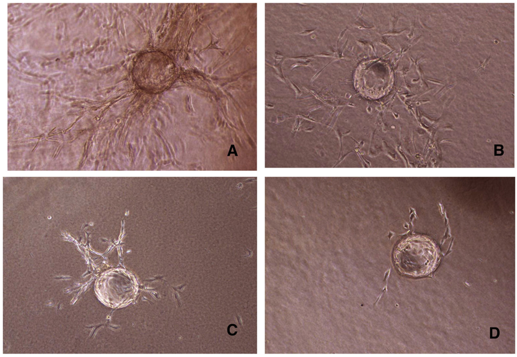
Fig. 3.
Formation of pseudo-capillaries from HBMEC. Cytodex microcarriers were coated with HBMEC, and then embedded in fibrin gel. Incubation was carried out for 2 days. Pseudo-capillaries sprouting were monitored by an inverted microscope at 20× magnification, using an ocular grid. Representative pictures of capillaries formation after FGF-2 stimulation (A) or after co-incubation with Aβ peptides (E22G, WT and E22Q; B, C, D, respectively) are shown.

