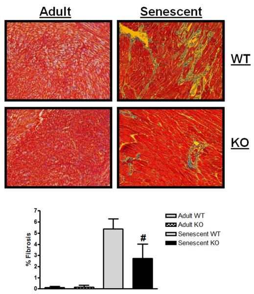
Figure 6. Senescent myostatin KO mice display less cardiac fibrosis.
Representative 20X images from Masson's trichrome stained heart sections from adult and senescent WT and KO mice are displayed with percent fibrosis data compiled below [adult WT, KO, senescent WT, KO; n=3,3,4,4]. Data are presented as mean ± SEM. #p<0.05 vs age-matched WT.

