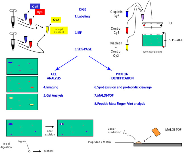
Figure 1. Work flow schematic for implementation of 2D-DIGE.
After homogenization proteins are labeled with up to 3 fluorescent dyes. The third dye, is usually used as an intragel standard for alignment of gels and quanfication of spots. Proteins are separated on 2D gels by charge (IEF) and molecular weight (SDS-PAGE). Analytical gels are scanned by a confocal-like scanner and gray scale images of each color are analyzed by software special for 2D-gels. Proteins of interest are excised robotically and cleaved, typically by trypsin, before laser irradiation. Ionized peptides accelerate though a magnetic field to the MALDI-TOF photomultiplier detector. Masses of peptides are determined with a precision of less than 1 atomic mass unit by measuring time of flight between the laser burst and the incidence of detection.

