
Figure 4. hNPC Cellular Graft Rejection.
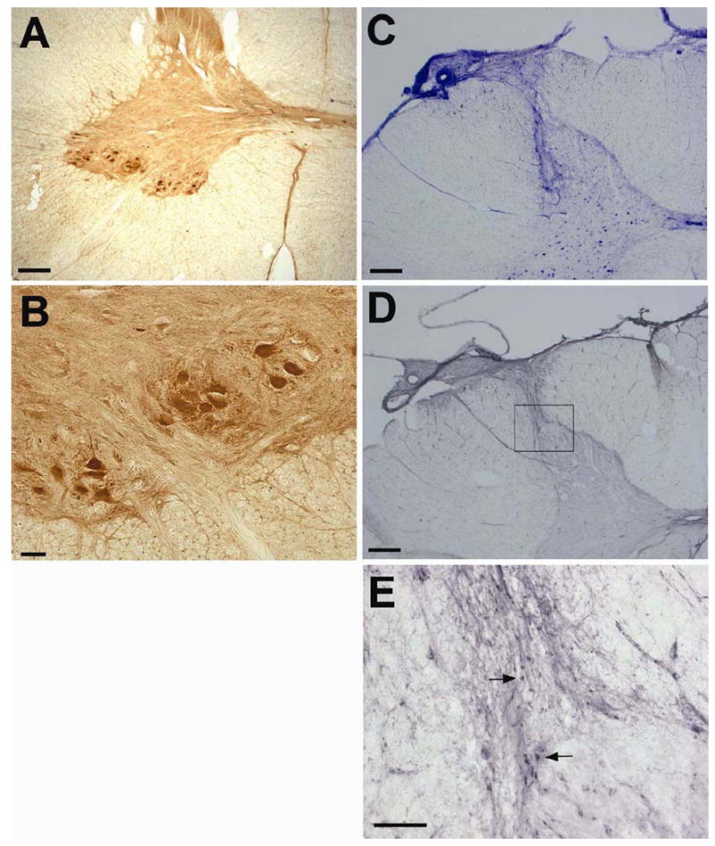
(A, B) Photomicrographs of choline acetyltransferase (ChAT) immunostaining in the section in the group 1 pig. Healthly ChAT positive motor neurons were found in the adjacent section of the injection site, indicating that the surgery procedure did not induce overt motor neuron damage. (C-E) Immunostaining for T cell marker CD8b shows an active cellular immune response in the core of the transplanted site. Animals in Group 4 received immunosuppression beginning 10 days prior to surgery with subsequent serial documentation of therapeutic CysA levels. While Nissl staining (C) showed the volumetric distribution of the cannula infusion track, significant T cell infiltration (designated by arrows in E) was detected in the injection core of the cervical spinal cord. Scale bars: 500 μm in A, C; 100 μm in B, D, E.