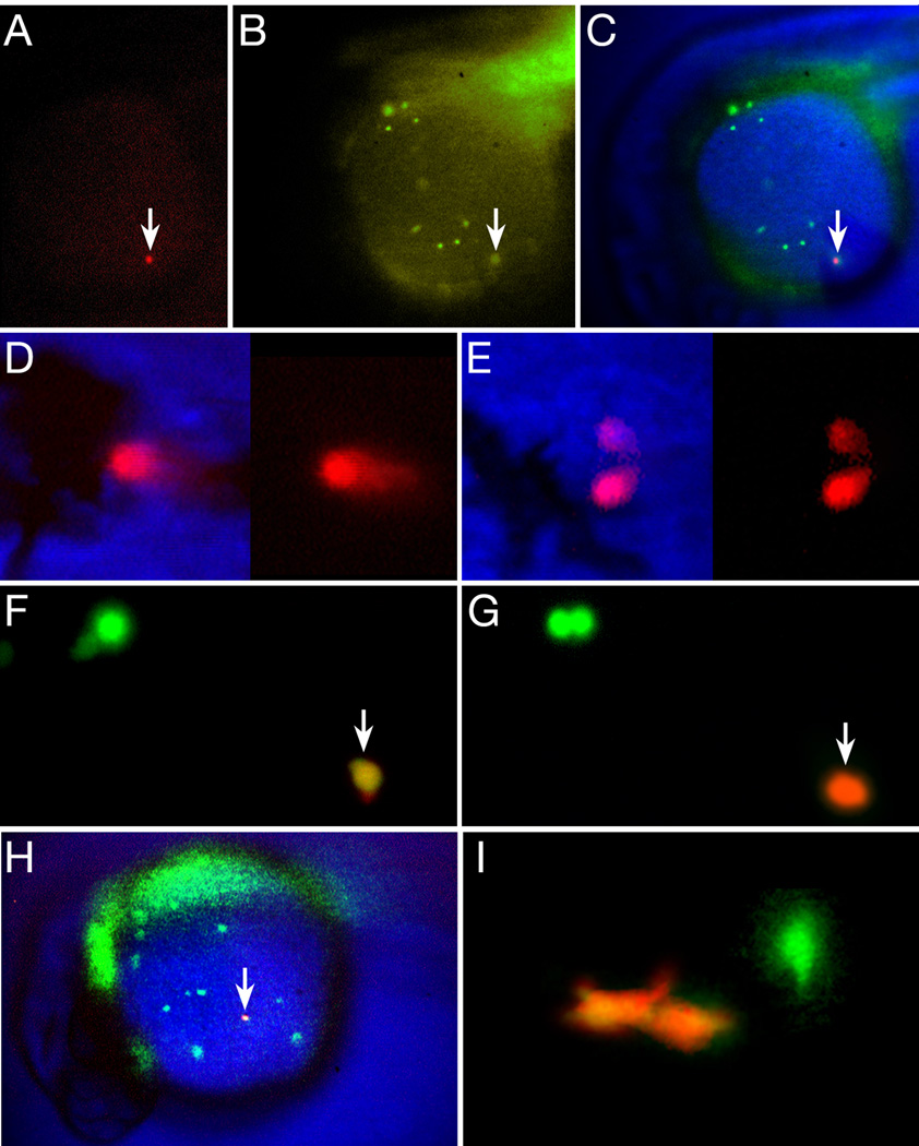
Figure 4.
Not all blood is committed to a unipotential lineage at 26 hours. (A–E) Single circulating blood cells give rise to neutrophil and erythrocyte progeny. (A–C) A newly photoconverted blood cell (arrow), within a ventral gastrula-derived clone. (A) Red wavelength, (B) green wavelength (note other blood cells) and (C) composite image. (D–E) The resulting red-labeled clone at 48 hours included (D) circulating erythrocytes and (E) 2 neutrophils attached to the lumen of a blood vessel. (F–I) Single macrophage cells give rise to macrophage progeny. (F–H) High magnification view of an individual macrophage (arrow) in the process of being photoconverted from green (F) to red (G). Nearby, another green-labeled macrophage (upper left panel) is preparing to divide. (H) Low magnification view of the entire Kaede clone showing the newly photoconverted macrophage (arrow) among other green-labeled macrophages and out of focus endoderm and endothelium. (I) The resulting red-labeled clone at 48 hours was solely 2 macrophages near a blood vessel. Another green-labeled macrophage is out of focus.

