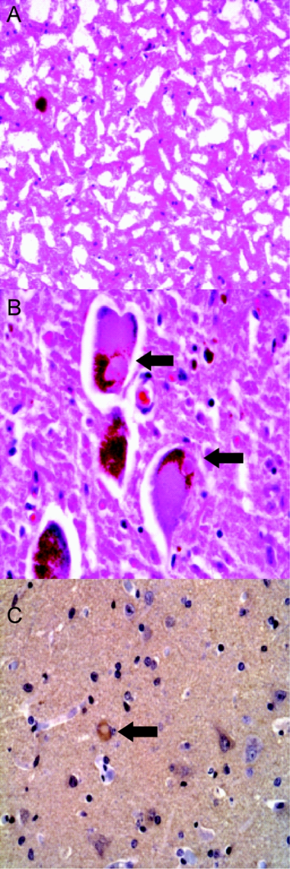
Figure 1 Parkinson disease neuropathology
Hematoxylin-eosin (H-E) stained frozen section of substantia nigra (SN) (A) and H-E stained paraffin section of locus ceruleus (LC) (B) showing severe pigmented neuron loss in SN and Lewy bodies in residual LC neurons (arrows). α-Synuclein immunostaining highlights rare immunoreactive neuron in frontal cortex (C).
