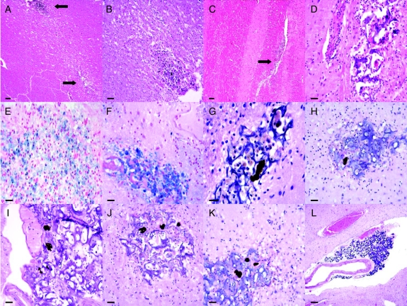
Figure 2 Representative tract lesions and associated findings
(A) Low power view of 2 tract sites, with upper lesion containing cluster of hemosiderin-containing cells shown in higher power in (B), and stained with Perl's iron in (E). (C) Linear tract lesion in internal capsule (arrow). (D) Tract focus containing matrix material without retinal pigment epithelial (RPE) cells. (F) Perl's iron stain of another tract site with matrix material and dense aggregate of likely hemosiderin-containing macrophages. (G–K) Hematoxylin-eosin stain of tract sites showing basophilic matrix material with representative RPE cell neuromelanin profiles. (L) Matrix material within Virchow-Robin space and abutting small artery in inferior putamen. Scale bar: A, C 200 μm; B 50 μm; D–F 30 μm; G 20 μm; H–K 30 μm; L 150 μm.

