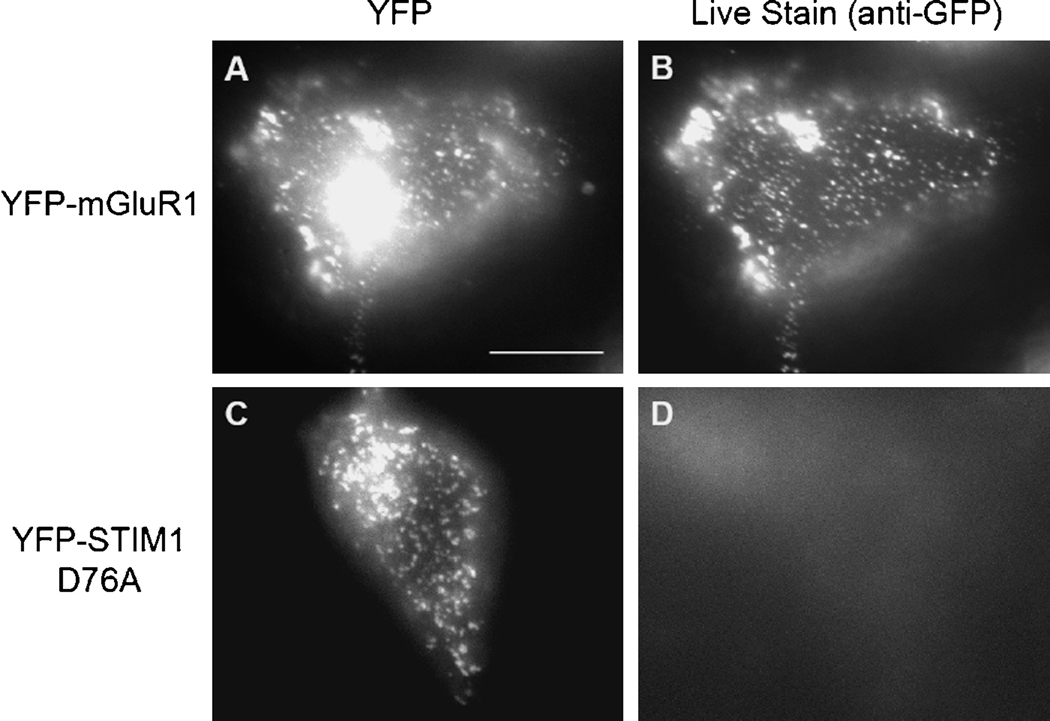
Fig. 1.
Majority of the YFP-STIM1 D76A punctae are not on the plasma membrane. HEK293 cells were transfected with YFPmGluR1 together with Homer1 and Shank3 to get the YFP-mGluR1 to the plasma membrane (A, B) or YFP-STIM1 D76A (C, D). A and C are intrinsic YFP signals. B and D are images of surface live staining with an anti- GFP rabbit polyclonal antibody. The intense signal in A is from Golgi. Scale bar, 10 µm.

