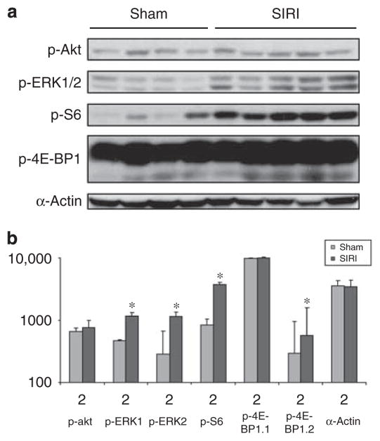
Figure 4. Analysis of signaling protein phosphorylation in ventricular tissue obtained from surgically induced renal injury (SIRI) or sham mice.
(a) Analysis of cardiac signaling protein phosphorylation in ventricular tissue obtained 8 weeks after SIRI (n=5) or sham surgery (n=4). Ventricular tissue was homogenized, protein lysates generated, and proteins were separated by SDS-PAGE followed by immunoblotting with primary antibodies directed against phosphorylated-Akt1/2 (p-Akt), phosphorylated-ERK1/2 (p-ERK1/2), phosphorylated-ribosomal S6 protein (p-S6), phosphorylated-4E-BP1, and α-actin as a loading control. (b) Scanned blot densitometry was performed with Image J (v1.24) software. *P<0.05 vs sham.

