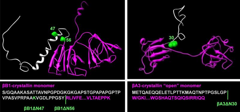
Figure 1. Three-dimensional models and sequences of βB1 and βA3 and N-terminal deletion variants.
The models of murine βB1 and human βA3 were generated by homology modeling as previously described (25). In βB1, the N-terminal extension consists of four regions where residues 1-28 are disordered, 29-41 and 43-47 form two short helical stretches and 48-57 a loop region connecting helix-2 and the main structural domain. The N-terminal extensions are colored grey, the sites of truncations, green, and the core structures, magenta. In βB1, S/G indicates mutation of Ser to Gly; in βA3, W/G indicates the mutation of Trp to Gly.

