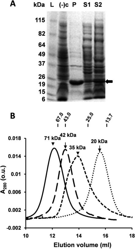
Figure 2. E. coli expression of β-crystallins and size fractionation of purified proteins.
(A) SDS-PAGE of βB1ΔN56; L, molecular weight marker; (−)c, BL21(DE3) competent cells only; P, pellet (insoluble fraction); S1, supernatant (soluble fraction) 1 mM IPTG, 37°C, 2 h; S2, supernatant, no IPTG, 21°C, 16 h (βB1ΔN56 protein indicated by black arrow); (B) Chromatography using Superdex 75 of βB1(short dash line), βB1ΔN56 (solid line), βA3 (dash line), and βA3ΔN30 (dotted line). Elution positions and molecular weights of protein standards are shown uppermost in figure.

