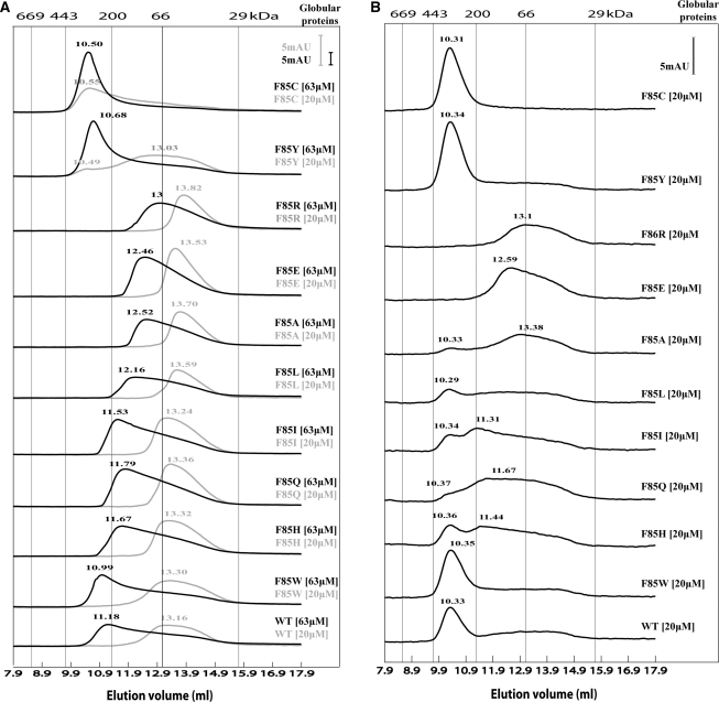
Figure 4.
Gel filtration profiles of the F85 variants at room temperature. (A) The elution profiles of the F85 variants chromatographed in the absence of nucleotide. Two concentrations were used for each F85 variant, low (20 μM light grey line) and high (63 μM black line). (B) The elution profiles of the F85 variants chromatographed in the presence of 0.4 mM ADP (using the lower 20 μM protein concentration). Standard globular proteins were used for calibration: thyroglobulin (669 kDa), apoferritin (443 kDa), β-amylase (200 kDa), bovine serum albumin (66 kDa) and carbonic anhydrase (29 kDa). Absorption unit (AU) on the scale corresponds to an A280 of 1.

