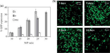
Figure 5.
(a) Percentage of MCEC expressing GFP 2 days post-transfection with peptides I, F or PEI. (b) Confocal images showing MCEC expressing GFP up to 14 days post-transfection with F/DNA complexes (N/P 20). Each image is a merger of several z-stack images acquired as the cell layers became overly confluent with the long incubation period. Cells also started to detach from the culture chambers after prolonged incubation, explaining the apparent reduction in cells expressing GFP on day 14.

