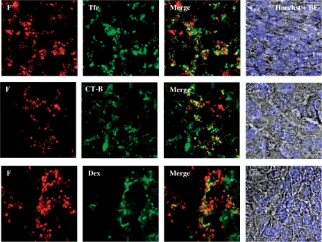
Figure 6.
Co-localization (yellow) of F peptide complexed with DNA (red) at N/P 20 with classical markers for specific endocytosis pathways like transferrin (Tfr, green) for clathrin-mediated endocytosis, cholera toxin B (CT-B, green) for caveolae/lipid raft-mediated endocytosis and dextran (Dex, false-coloured green) for macropinocytosis. Cells were all stained with the Hoechst nuclear dye (blue) and presented with the bright field (BF) images. Imaging was done 40–90 min post-exposure to peptide/DNA complexes.

